What is the Stroke Code?

Reading time: 3 min

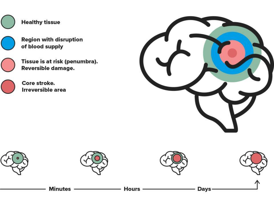
Of all the organs, the brain is the most sensitive to a lack of blood supply. Therefore, and in order to preserve brain function, it is vital to diagnose stroke early on and administer a specific treatment to re-establish blood flow and prevent further tissue damage.

Rapid intervention in the first hours will increase the chance of greater functional recovery while decreasing the sequelae (long-term after-effects) and mortality.

In Catalonia, a strategy called “Stroke Code” has been developed to ensure patients receive help as soon as possible. “Stroke Code” is an emergency protocol that quickly identifies, reports and transfers patients with a suspected stroke to their reference hospital, where they then undergo an accurate and early diagnosis before receiving the most appropriate treatment for each case.

Whenever a stroke is suspected, the doctor must assess the symptoms very accurately and rule out any other conditions which can mimic stroke, for example a decrease in blood sugar (hypoglycaemia).

Emergency tests are used to diagnose the stroke and determine whether it is an infarction or haemorrhage. The aim is to guide initial treatment and limit the after-effects.

Magnetic Resonance Imaging or MRI machine

The main tools used for emergency diagnosis of stroke are neuro-imaging techniques, such as computed tomography (brain CT scan) or magnetic resonance imaging (brain MRI scan).

Cerebral CT angiography or computed tomography angiography

Tests that offer a direct visualisation of blood vessels, such as computed tomography angiography (CTA), are usually performed. An angiography provides a detailed image of flow through the body’s blood vessels. A CTA scan of the brain can locate a blockage or check whether blood is reaching all areas of the brain correctly.

Once stroke is diagnosed, most cases generally require additional tests to determine the cause. The main objective is to guide post-stroke prevention treatment and prevent recurrence, but the treatment will vary in function of the cause:

Ultrasound on a monitor

Doppler ultrasound is a type of ultrasound used to determine the volume, rate and thickness of blood flowing through arteries and veins at a given moment. The technique can also be used to detect alterations (e.g., arteriosclerosis plaques) in the major arteries, principally those in the neck, carrying blood from the heart to the brain.

Monitor with an echocardiogram

Echocardiography is another commonly practised test that is used to study any anatomical changes in the heart.

Electrocardiogram monitor

Cardiac rhythm studies, such as an electrocardiogram or a Holter test, may also be appropriate.

Blood collection tube

Some of the indicators measured in laboratory tests include sugar and cholesterol levels, plus counts of platelets and other types of blood cell which can induce thrombosis. More specific laboratory tests are sometimes conducted in search of hidden tumours, coagulation disorders predisposing thrombosis or genetic alterations that may increase the chance of stroke, amongst others.

Substantiated information by:

Antonia Fernández
Arturo Renú Jornet
Xabier Urra Nuin
Ángel Chamorro Sanchez

Published: 20 February 2018
Updated: 27 December 2022

The donations that can be done through this webpage are exclusively for the benefit of Hospital Clínic of Barcelona through Fundació Clínic per a la Recerca Biomèdica and not for BBVA Foundation, entity that collaborates with the project of PortalClínic.

Subscribe

Receive the latest updates related to this content.

Thank you for subscribing!

If this is the first time you subscribe you will receive a confirmation email, check your inbox

An error occurred and we were unable to send your data, please try again later.