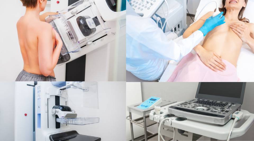
A mammogram is a high-definition, very low dose X-ray of the breast to detect breast cancer or other breast diseases, and can find early stage lesions.
Meanwhile, a breast ultrasound scan is a scanning technique that uses ultrasound waves. It is recommended especially for women under 35 years of age as an initial diagnostic test and sometimes for women over that age in addition to mammography. After the age of 35, mammograms are recommended, since they are more accurate for detecting breast cancer, as microcalcifications can be observed, which may be the first signs of the disease. It should be remembered that the risk of developing breast cancer increases with age.
Mammograms:
- These are X-rays.
- They are performed standing up with a device called a mammograph which requires moderate compression of the breast.
- No special preparation is necessary for the test, but the mammogram should be done the week after menstruation.
Breast ultrasound scans:
- These use a probe impregnated with a gel that facilitates movement.
- Ultrasound waves, not X-rays, are used, which reflect off the explored area, specifically the breast, and are converted into images.
- The result is not as reproducible as mammography, as it depends on the operator obtaining the images. However, an advantage is that it does not require the breast to be compressed.
- No prior preparation is needed for breast ultrasound.
Currently, most mammography machines are digital and have the advantage of better x-rays being done at lower radiation doses. Some devices now come with new 3D or tomosynthesis technology, which provides tomographic images of the breast and helps to differentiate certain lesions and pinpoint their location. Lighter or darker areas can be seen on the mammogram, according to the breast density. Before the test, the health professional should be informed of pregnancy, lactation, a recent operation, if the patient has received a blow or if she is carrying a breast prosthesis. During the test, all metallic objects have to be removed and no body creams or deodorants can be applied, so as not to interfere with the image. It is also important to tell the doctor about the use of hormones and any family or personal history of cancer. If you notice any discomfort during the test, tell your healthcare professional.
The person who interprets the results is a doctor specialising in radiology, with specific breast diagnostic training. In some cases, a mammography is performed alongside a breast ultrasound if extra examinations are needed. This does not mean something abnormal has been seen, but rather that other images are needed.


