Capsule endoscopy is the first imaging test capable of examining the small intestine in detail, using a tiny camera about the size of a pill. This examination has transformed the diagnosis of patients with suspected small bowel disease, as it provides earlier and more efficient diagnosis, reducing healthcare costs.

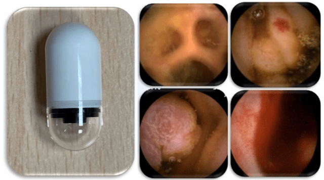
Endoscopic capsule device (left) and images of the small intestine (right).
Patient preparation and carrying out the test are very simple
Before the examination, the patient is recommended to prepare for it by taking a simple laxative and, sometimes, a medication that increases intestinal movement, to improve the transit of the capsule.
The examination is performed on an outpatient basis, that is, without hospital admission. The patient is fitted with a belt with sensors to capture the images from the capsule and this, in turn, is connected to a recorder. After taking the capsule, the patient goes home and resumes a normal life. 2 hours after ingestion, the patient can resume taking his usual medication; then, 4 hours after, he can have a light meal. Moderate physical activity should be maintained. After 8 or 9 hours, the belt is removed and the images are downloaded from the recorder in the endoscopy consulting room.
An examination for the detection of diseases of the small intestine
This minimally invasive technique has proven to be highly sensitive in detecting lesions of the small intestine; as good as or better than other imaging tests. The main contraindication for capsule endoscopy is intestinal obstruction.
According to current clinical guidelines, capsule endoscopy is the examination of choice for the study of mid-gastrointestinal bleeding. This condition is seen when persistent and recurrent bleeding of an unknown origin occurs during a conventional endoscopic evaluation of the digestive tract (gastroscopy and colonoscopy). It is also indicated for monitoring and diagnosing inflammatory bowel disease or assessing intestinal lesions caused by anti-inflammatory drugs, such as ibuprofen. The technique has also been shown to be effective for diagnosing small intestine tumours or celiac disease that does not improve with the elimination of gluten from the diet.
The device emits the images with signals similar to radio transmission
This new pill-sized 11 x 26 mm endoscopic device has a built-in camera, which records images of its entire journey through the intestine. It emits the images through a transmitter that sends them to sensors located in the abdomen, similar to radio transmission. The sensors are incorporated into a belt placed around the patient's waist. These send the signals to a recorder attached to the belt that the patient wears for the duration of the test.
After ingesting the capsule, it moves through the digestive system with the natural movements of the intestine itself, sending 2-6 photos per second during 9-12 hours of recording. The images are subsequently processed and viewed like a film.
The device is expelled spontaneously from the rectum in the stool. This expulsion can vary depending on the intestinal rhythm of each patient. If it has not been expelled after two weeks, the doctor should be consulted, as it may have been retained in the digestive tract.
In fact, the main complication of this test is retention of the capsule in the intestine. This occurs in 1-5% of patients and is slightly higher in patients with Crohn's disease. If this occurs, it is usually asymptomatic and the capsule can be expelled spontaneously with the help of medication. If not, it can be removed by specific endoscopic examination (deep enteroscopy). This complication can be prevented by using an absorbable capsule that confirms the patency of the digestive tract.
Pregnancy is a relative contraindication due to the existence of few published studies. For patients with swallowing problems, the examination can be performed by placing the capsule in the intestine endoscopically using a specific device.
Capsule endoscopy makes it possible to diagnose diseases of the small intestine early and cost efficiently. Despite this, if the disease is suspected to occur in another part of the digestive tract - such as the oesophagus, stomach or colon - conventional endoscopic tests, gastroscopy or colonoscopy, are still the tests of choice.
INFORMATION DOCUMENTED BY:
Begoña González Suárez, Specialist in Gastroenterology and Digestive Endoscopy in the Digestive Endoscopy Section of the Gastroenterology Service at the Clínic Barcelona Hospital and associate professor at the University of Barcelona.


