The study, published in the journal Insights into Imaging (Q1), shows that integrating this tool into the workflow could improve the efficiency of lesion classification and reduce the workload of radiology specialists.
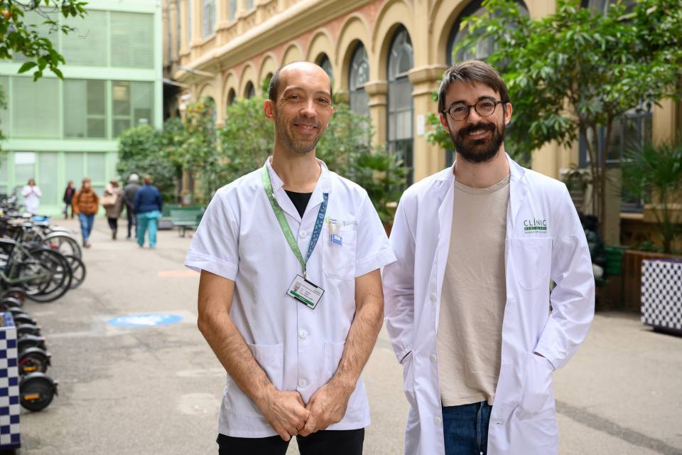
The study was coordinated by Dr Jordi Rimola and Dr Mario Matute, radiologists in the Liver Cancer Unit at the Clínic Barcelona and researchers in the IDIBAPS Liver Cancer (BCLC) group. It received technical support from the IT Department of the Spanish Association for the Study of the Liver (AEEH).
Integrating AI into radiology
Natural language processing (NLP) is a field of artificial intelligence that focuses on developing algorithms and models for machine learning systems to interpret human language. Together with large language models (LLMs), these tools have aroused great interest among the general public, and specifically in the field of medicine as well.
At present, most radiology reports are written in a free-text format, which makes it difficult to extract structured information for research and clinical decision-making. Thus, both natural language processing and LLMs models have emerged as promising tools for addressing this challenge.
In the field of radiology, several recent studies have demonstrated the usefulness of LLM in report standardization and the extraction of structured and quantitative information from free-text radiology reports. “Moreover, unlike older NLP models, the new LLMs are trained on unlabelled data and can be applied to complex tasks with little specific information”, says Mario Matute.
LiverAI: A model for automated data extraction
LiverAI, developed on GPT-4 architecture, was trained to categorize liver lesions according to the LI-RADS v2018 system, used to assess the risk of malignancy in patients at risk of developing liver cancer. The tool was tested with a set of real data on liver lesions in patients with cirrhosis and compared with the classification made by expert radiologists.
Results and clinical implications
The study showed that LiverAI achieves a moderate level of concordance with the radiologists’ assessment, but, when used as a complementary tool in the workflow, it can improve the efficiency of the process by up to 60%. In particular, the model demonstrated a high sensitivity for identifying lesions that are probably malignant, a fact that suggests that it could be used as a triage tool to optimize the work of professionals.
“The study is a proof of concept, but it offers new perspectives on the potential applications of language models, presenting a real example of how these tools could be integrated locally to optimize data collection in a research context”, explains Jordi Rimola.
This research represents a step forward in the integration of artificial intelligence into the field of radiology, and opens the door to future applications for the automation of data extraction in biomedical research. In this field of research, the liver oncology group recently incorporated an expert in artificial intelligence supported by a Marie Cure grant, which will allow research to advance in this area.
Study reference:
Matute-González M, Darnell A, Comas-Cufí M, Pazó J, Soler A, Saborido B, Mauro E, Turnes J, Forner A, Reig M, Rimola J. Utilizing a domain-specific large language model for LI-RADS v2018 categorization of free-text MRI reports: a feasibility study. Insights Imaging. 2024 Nov 22;15(1):280. doi: 10.1186/s13244-024-01850-1.


