18 December 2025
What is PET-CT?
Computed Tomography combined with Positron Emission Tomography or PET-CT is a nuclear medicine diagnostic test that combines two different techniques to provide detailed information about the human body, offering a hybrid view of the metabolism (with Positron Emission Tomography, PET) and the anatomy (with Computed Tomography, CT).
This combination helps healthcare professionals identify potential health problems, such as tumours or other disorders, and determine their precise location within the body. It is a valuable tool for diagnosis and treatment planning for many medical conditions.
PET-CT imaging is used in a wide variety of diseases, with a high volume of its use being in the oncology field of healthcare. PET is a minimally invasive technique via intravenous administration. It is safe and can be repeated as many times as necessary.
How does it work?
A radioactive substance (harmless to the body), bonded to a specific molecule (known as a radiotracer or radiopharmaceutical), is administered to the patient and then travels to the organ or tissue to be studied. The radiopharmaceutical then decays and emits positrons. These interact with electrons in our body and produce gamma rays, which are detected by a special camera called a PET tomograph. The tomograph produces an image of the distribution of the radiopharmaceutical within the body.
The PET tomograph is then combined with the CT technique to provide detailed anatomical structural images of organs and tissues. This provides information about the exact location of the areas highlighted by the PET.
PET-CT offers:
- Better resolution. Locating areas with abnormal metabolic activity more accurately.
- More sensitivity. It can detect a smaller amount of radiopharmaceutical.
- Shorter image acquisition time when compared to SPECT/CT, another nuclear medicine diagnostic test.
What radiopharmaceuticals are used?
The radiopharmaceuticals used in PET have specific targets which provide more accurate images, and therefore more accurate diagnosis and better treatment planning.
The radiopharmaceutical used most is fluorodeoxyglucose (18F-FDG) as it concentrates in areas of high glucose consumption and thus reflects the metabolic activity of cancer cells. A number of other radiopharmaceuticals are used for various clinical applications in PET, such as 68Ga-DOTA-Octreotide (DOTATOC) for patients with neuroendocrine tumours and to detect and monitor liver and other metastases; 18F-choline (fluorocholine)/18F-PSMA, used primarily in prostate cancer imaging; and 18F-flutemetamol/18F-florbetaben, used to detect brain amyloid plaque in patients suspected of having Alzheimer's disease or other neurodegenerative disorders.
When does a PET-CT need to be performed?
A PET-CT may be required to assist in diagnosis, treatment planning and evaluation of response to treatment in several clinical situations. A decision to perform a PET-CT depends on the indications of health professionals and the specific clinical situation of the patient they are treating. Not all patients require a PET-CT and its potential use is evaluated according to an individual’s symptoms, the results of other tests and the overall clinical picture.
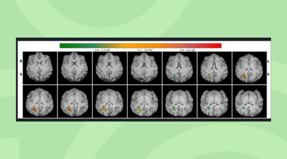
For example, one of the most common applications of this technique is to determine the best therapy for oncological diseases by detecting tumours, determining their extent and assessing the response to treatment. In cardiovascular diseases, it can evaluate blood flow in the heart and detect problems such as coronary artery disease. In neurological diseases, such as dementia or epilepsy, it can provide information about the metabolic activity of the brain. In inflammatory diseases, it can be useful for detecting and monitoring inflammatory processes, such as temporal arteritis or other autoimmune disorders. It can also help locate and characterise infections in various parts of the body.
How should I prepare for the procedure?
Preparation for PET-CT varies with the specific type of examination and the medical condition being evaluated. Some general guidelines however are:
- Inform the professional about any medical condition, allergy you have or medication you are taking, especially if you have diabetes.
- It is important to inform health professionals if you have diabetes and to follow their instructions before undergoing the test. You should also have diabetes medication with you.
- Wear comfortable clothing, preferably without metal zips or buttons, as they can interfere with the image. All metallic objects, such as jewellery, watches or coins, must be removed.
- Drink plenty of fluids before and after the test, as this helps clear the radiopharmaceutical from your system more quickly.
- You need to fast beforehand, except for unusual PET radiopharmaceuticals indicated by the healthcare professional.
- Avoid caffeine, nicotine and strenuous exercise for at least 24 hours before the procedure, as they can affect the results.
- If you are breastfeeding, pregnant or suspect you may be pregnant, inform your healthcare professional or nuclear medicine nurse beforehand, as some types of nuclear medicine imaging may not be recommended during pregnancy or breastfeeding.
- Arrive early for the appointment and leave enough time for the procedure and any additional paperwork that may be necessary.
- You may have to stay in the imaging centre after injection, to give time for absorption of the radiopharmaceutical and for taking the images at the optimal time.
- Close contact with pregnant women or children must be avoided for as long as indicated by the nuclear medicine nursing staff.
How is it performed?
The specific steps for performing a PET test vary with the type of radiopharmaceutical and medical condition, but the general steps are as follows:
- The radiopharmaceutical is administered to the patient intravenously.
- The patient must wait in the centre for the time indicated by the nursing staff so that the radiopharmaceutical is distributed throughout the body.
- After the waiting period, the patient is asked to lie still on a bed. A CT image is taken, followed by a PET image of the area of interest.
- In some types of PET-CT, the patient is asked to change position during the process to obtain images of other areas of the body.
- Once the test is finished, the patient may be asked to wait while the images are reviewed by the nuclear medicine specialist, to check they can be used for assessment.
- The radiopharmaceutical administered breaks down naturally after the examination, and is eliminated from the body in the urine and faeces. The patient is advised to drink fluids to help clear the radiopharmaceutical from the system more quickly.
Where is the test performed?
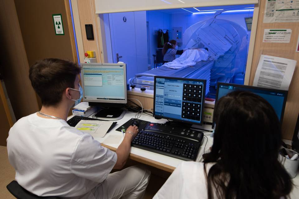
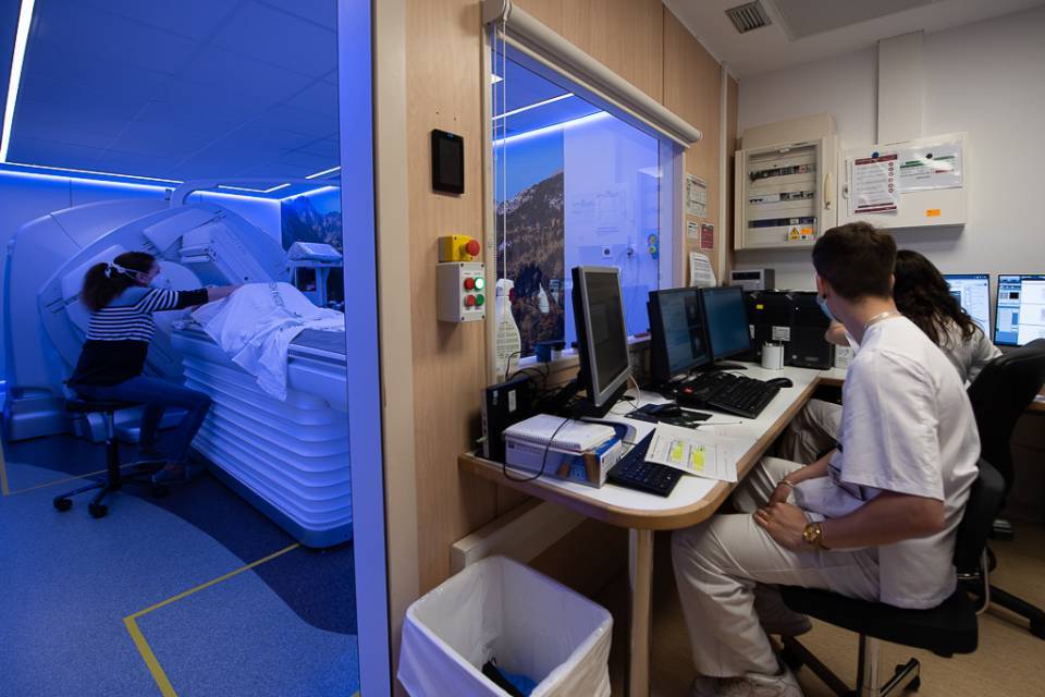
PET-CT is performed in the nuclear medicine unit of medical centres.
Because PET-CT imaging involves the use of radioactive materials, strict safety procedures are followed to minimise radiation exposure for patients, healthcare workers and the general public.
Who performs the test?
The PET scan is performed by a qualified team of nuclear medicine specialists. The team includes:
- A nuclear medicine specialist. Responsible for interpreting the examination results from the images obtained and determining the best treatment.
- Radiopharmacist. Responsible for radiopharmaceutical management.
- Nuclear medicine nurse. They assess the patient beforehand and administer the radiopharmaceutical based on the test indicated by the specialist.
- Diagnostic imaging technician. Responsible for operating the PET-CT tomograph and ensuring patient safety during the examination.
- Biomedical engineer Creates and supervises the image acquisition, reconstruction and processing features.
- Radiophysicist. A specialist who ensures that the PET-CT scanner is working correctly and that the radiation dose administered to the patient is safe and appropriate.
All team members are trained professionals who work together to ensure the examination is performed safely and accurately and that the patient receives the best care possible.
How long does the test last?
The time taken for the PET scan varies according to the type performed. Some procedures may take 1 to 3 hours.
For example, a whole-body PET-CT with 18F-FDG usually takes 90 minutes to complete, during which time the patient is inside the machine for only 25 minutes.
You should arrive on time for your appointment and follow the preparation instructions provided by your healthcare team to ensure the examination is performed efficiently and accurately.
What will I feel during the test?
Most patients do not experience any discomfort during a PET-CT. However, some patients may experience some of the following sensations:
- Discomfort from the injection. The patient may feel a little discomfort or pain at the injection site.
- Heat or cold. Patients may feel a sensation of warmth or cold as the radiopharmaceutical is absorbed.
- Nausea. Some patients may experience mild nausea or a metallic taste in the mouth as a side effect of the radiopharmaceutical.
- Need to urinate. Patients may experience the need to urinate more often, as the radiopharmaceutical is removed from the body.
- Claustrophobia. Because they have to remain still in a confined space during imaging, some patients can feel claustrophobic or anxious.
You should inform a healthcare team member if you experience any discomfort or side effects during the scan. Staff can provide additional information and support to help make the scan as comfortable as possible.
What are possible complications?
PET-CT is considered a safe, non-invasive imaging technique. However, there are potential complications associated with the use of radiopharmaceuticals:
- Allergic reaction. In very rare cases, patients may experience an allergic reaction to the radiopharmaceutical injection, causing symptoms such as hives or itching. If iodinated contrast is administered, allergic reactions may appear, much as they might if a CT scan is performed.
- Radiation exposure. The dose administered in a PET procedure is very low and the risk of long-term side effects is generally low, so it does not interfere with normal life.
- Pregnancy and lactation. Pregnant or breastfeeding women should inform their healthcare professional before undergoing a PET scan, so as to minimise the radiation received by the foetus or infant.
You should discuss any potential risks or concerns with the specialist before undergoing a PET-CT scan. The professional will be able to provide additional information and help resolve any questions about the procedure.
Related contents
Substantiated information by:

Published: 23 February 2024
Updated: 23 February 2024
Subscribe
Receive the latest updates related to this content.
Thank you for subscribing!
If this is the first time you subscribe you will receive a confirmation email, check your inbox


