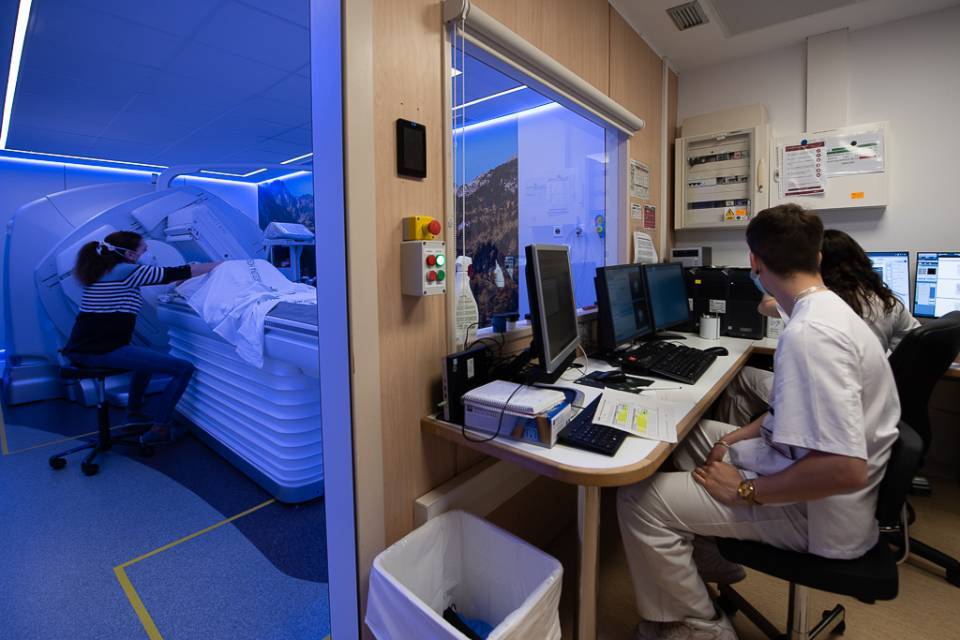
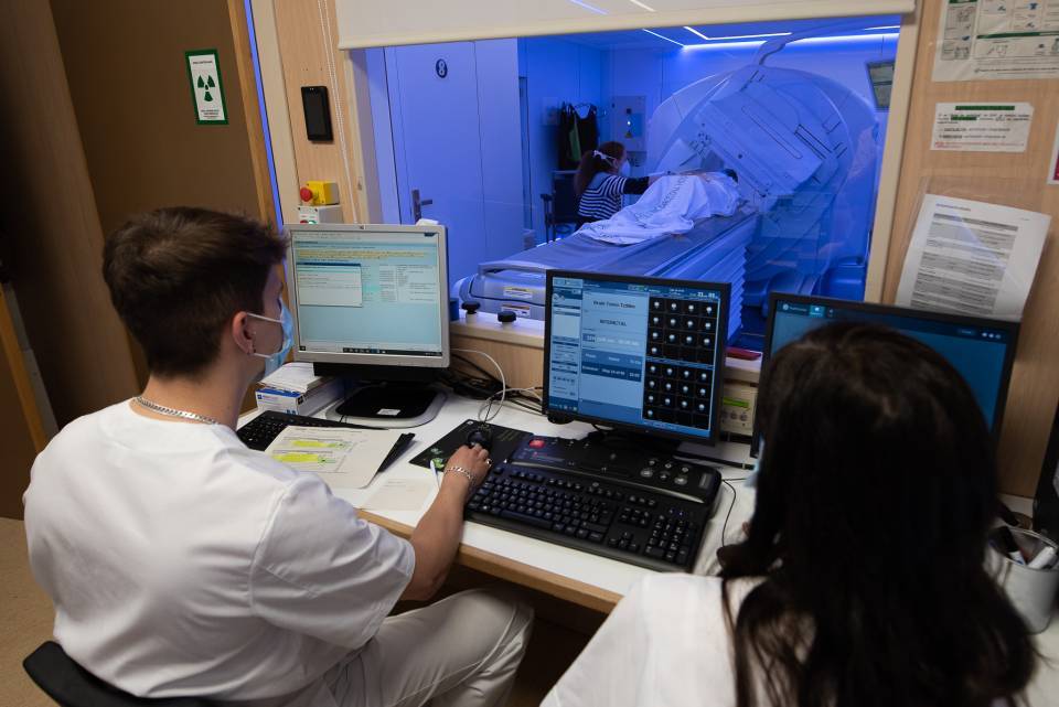
The Hospital Clínic de Barcelona has incorporated a new gamma camera (digital SPECT/CT) for diagnosis using radionuclide imaging. Up until now, there was no technology with these characteristics in operation in Spain.
The new gamma camera implements digital direct conversion detectors, which identify the location of the scintillation event generated, without the need for rough calculations. Moreover, it eliminates the analogue noise that is produced, avoiding any loss of signal. All this results in improved performance and clinical utility.
The gamma camera is a device commonly used in nuclear medicine, and allows us to study a wide variety of diseases. The device detects the gamma radiation produced after a radioactive tracer is administered to the patient (usually by intravenous injection) and generates an image that provides information on an organ's activity at a molecular level. The scintigraphic image provides valuable morphological and functional information about a specific organ or tissue. This technique is applied in the study of a wide variety of systems such as the osteoarticular, genitourinary, digestive, cardiovascular, respiratory and endocrine systems and the brain.
This type of digital technology will allow for a significant reduction in the time required for imaging and, therefore, increase the number of examinations by up to 30%, shortening our patient waiting lists. Moreover, the improvement in the resolution of the images will also help us to increase the diagnostic accuracy of small lesions, which could go undetected with analogue technology.
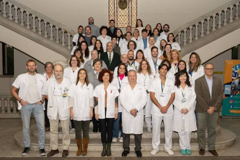
According to Dr. David Fuster, head of the nuclear medicine service at the Hospital Clínic’s Diagnostic Imaging Centre (CDIC), “the incorporation of this new digital molecular imaging technology will allow for better diagnoses and improve the characterization of lesions.” “With the incorporation of the new gamma camera, we reaffirm our commitment to the use of the most cutting-edge, patient-oriented technology,” he concludes.


