- What is it?
- Team and structure
What is a Hysteroscopy?
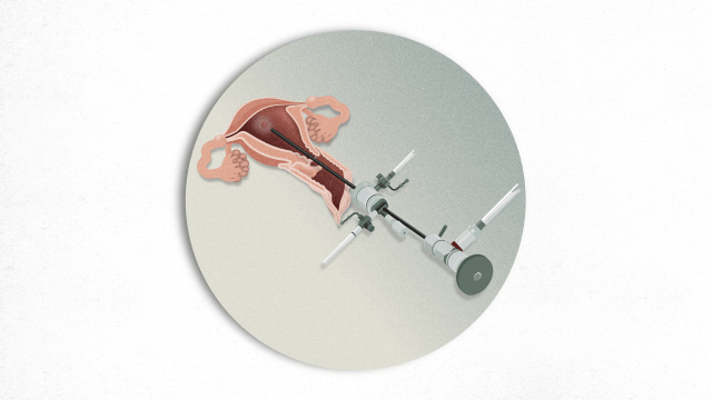
Hysteroscopy is an endoscopic procedure for direct visualisation of the vagina, cervix and uterine cavity using an optical device (hysteroscope) inserted through the vagina and endocervical canal until it reaches the uterine cavity.
Obtaining a tissue sample (biopsy) for further analysis may sometimes be necessary during the hysteroscopy. It is a simple and safe procedure with very few risks and complications.
What does it consist of?
It is performed by inserting a small endoscope (an optical device) through the vagina and the cervix. To gain the best view of the uterine cavity possible, it is distended with a saline solution.

What is it for?
It allows us to diagnose uterine cavity conditions. It has many indications, although the most common are to study abnormal bleeding, conditions of the endometrium (a layer of tissue that lines the uterus) or myometrium (the outer muscular layer of the uterus wall), infertility or postoperative control of uterine surgery.
The most common findings are:
- Endometrial polyps (growths inside the uterus).
- Intracavitary fibromas or myomas (non-cancerous tumours of the uterus).
- Endometrial disease such as endometrial hyperplasia (excessive growth of endometrial cells) or endometrial cancer.
It is also indicated in many cases for the study of reproductive problems: suspicious alterations of the uterine cavity identified in gynaecological ultrasound or other imaging techniques such as uterine malformations or intrauterine adhesions. It is also used to diagnose subtle endometrial changes such as chronic endometritis (chronic inflammation of the endometrial lining of the uterus).
It is also indicated to assess the uterine cavity in patients who have had previous uterine surgery. A hysteroscopy can also be used to remove an intrauterine device (IUD) from the endometrial cavity.
How it is performed and how long does it last?
The patient lies on her back and places her legs on the table's stirrups. The hysteroscope is inserted through the vagina and cervical canal using saline to distend it and the interior of the uterine cavity is inspected. The video camera allows the images to be viewed via a television monitor. It is performed in an outpatient consultation, without the need for anaesthesia and lasts approximately 2-3 minutes.
How do you prepare for it?
No special preparation is necessary. Performing it in the first half of the menstrual cycle is recommended (it cannot be done during menstruation).
Special attention
Patients with a history of infertility, pelvic inflammatory disease or hydrosalpinx (accumulation of fluid in one or both fallopian tubes that can cause infertility problems) require an antibiotic treatment prior to the procedure.
In some cases, an antibiotic treatment may be required for patients with endocarditis (infection of the endocardium, which is the inner lining of the heart valves and chambers) or those who have mechanical valves.
Who performs the test?
A specialist healthcare professional will guide, advice and monitor the patient throughout the test.
What will I feel during the test?
After the examination, there may be mild discomfort similar to mild menstrual pain. You may also experience a small amount of blood loss for several days after the procedure.
At the end of the test, you can carry out daily activities as normal. However, sexual intercourse or submerging yourself in baths or pools is not recommended until 24 hours after the test.
Related contents
Substantiated information by:

Published: 19 October 2020
Updated: 31 January 2025
Subscribe
Receive the latest updates related to this content.
Thank you for subscribing!
If this is the first time you subscribe you will receive a confirmation email, check your inbox