5 February 2020
- What is it?
- Causes and risk factors
- Symptoms
- Tests and diagnosis
- Treatment
- Disease evolution
- Research lines
- Frequently Asked Questions
-
La enfermedad en el Clínic
-
Equipo y estructura
Tests and diagnosis of Uveitis
The ophthalmologist may perform a variety of successive tests to diagnose uveitis:

Visual acuity. The most important functional parameter is visual acuity. It may decrease due to corneal or crystalline lens opacity, inflammation of the anterior chamber or the vitreous chamber, altered function of the retina or optic nerve, or due to the onset of complications, such as macular oedema. Visual acuity should be measured at each visit and can be used to monitor the clinical evolution of uveitis and the response to treatment and is therefore extremely useful when it comes to making therapeutic decisions. It is quick and easy to assess and provides a lot of information.

Intraocular pressure in both eyes. The most accurate test is called Goldmann applanation tonometry and involves instilling a fluorescein eyewash, often yellowish in colour, with a short-acting local anaesthetic. It is important to evaluate the intraocular pressure because some types of uveitis are associated with ocular hypertension. Inflammatory ocular hypertension (uveitic glaucoma) entails complicated treatment as it has a negative long-term effect on visual acuity.

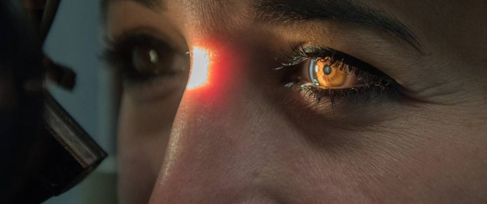
Slit lamp examination. During the ophthalmological examination the doctor observes the eye with a special microscope called a slit lamp. In cases of uveitis the specialist may observe the presence of inflammatory cells floating in the intraocular fluids and can determine the degree and type of inflammation. To examine the back of the eye, where the retina and optic nerve are located, the ophthalmologist will place some pupil dilating eye drops, called mydriatics, such as tropicamide or cyclopentolate. These drops can temporarily blur your vision.

Retinography. A retinography is a photo of the eye fundus used to examine the retina, optic nerve, vascular tree and macula. Thanks to new wide-field imaging techniques, ophthalmologists can now observe a 200° extension of the retina and even up to the peripheral retina.
Slit lamp
The slit lamp examination must be performed by an ophthalmologist to assess the condition of the anterior segment of the eye. Inflamed eyes often present aggregates of inflammatory cells on the posterior side of the cornea (subendothelial keratic precipitates). The presence of inflammatory cells (Tyndall effect) and proteins (aqueous flare) in the aqueous humour is indicative of a broken blood-aqueous barrier and can be indirectly correlated to the degree of inflammation. Inflammation is graded from 0 to 4+ according to the intensity.
When there are a lot of inflammatory cells in the anterior chamber they form a fluid deposit known as a level of hypopyon (pus). One of the common complications associated with uveitis is the formation of anterior or posterior synechiae (when the iris adheres to the iridocorneal angle or crystalline lens, respectively). If there is inflammation in the intermediate or posterior segments of the eye, then inflammatory cells may be observed in the vitreous, a phenomenon known as vitritis or vitreous Tyndall.
The degree of vitritis can also be graded from 0 to 4+. An eye fundus examination is essential in all inflammatory processes of the eye and is performed with either a direct or indirect ophthalmoscope and the aid of lenses. The exam must be done by trained specialists and is usually carried out by an ophthalmologist.
Ophthalmoscopy helps differentiate lesions in the retina or choroid, such as focal areas of inflammation or necrosis, or signs of retinal vasculitis, and determine whether or not the optic nerve is affected. Depending on the location of the inflammatory focus the condition can be classified as retinitis (in the retina), choroiditis (choroid), chorioretinitis (choroid + retina) or neuroretinitis (optic nerve + retina).
In function of the results, additional tests may be required:

Perimetry. A perimetry, campimetry or visual field test is an examination used to assess any alterations in the patient’s visual field. The visual field is the total space the eye can capture while focusing on a central point. Ophthalmologists can use this test to detect any type of peripheral vision loss and they also obtain a map of this loss which guides them in the diagnosis of certain pathologies (glaucoma, diseases of the retina, optic tract lesions, etc.).
Patients do not need to do anything to prepare for this type of test, which is painless and does not have any contraindications.

Optical coherence tomography (OCT). OCT is a complementary test used to determine whether the retina is working correctly and if there is any intra/subretinal fluid present.
OCT angiography is an advanced imaging test that examines the retina's blood vessels without the need for contrast. It helps detect neovascular membranes or hypoperfusion (decreased blood flow) in some cases of uveitis.

Fluorescein angiography (FAG). Fluorescein angiography examines blood flow in the posterior portion of the eye. A contrast agent is injected intravenously and circulates through vessels in the retina, thus affording a real-time evaluation of the retina’s vascular tree. In cases of uveitis, the fluorescein dye can be seen leaking from the vessels because they are inflamed. Whereas in retinal vascular diseases, including vasculitis, FAG reveals whether there are any areas of the retina with poor blood flow (ischaemia) or which have even developed new vessels.

Indocyanine green (IGG). Is an imaging test performed after FAG, which uses a different type of dye. It allows visualization of the deepest layer (the choroid) thanks to the chemical characteristics that differentiate it from FAG. It is especially useful in uveitis affecting the choroid stroma, such as Voght-Koyanagi-Harada, Birdshot disease, or ocular sarcoidosis, or in infectious diseases such as tuberculosis.
This test helps us detect choroidal inflammation that sometimes goes unnoticed in the fundus or cannot be detected with other tests.
Multidisciplinary diagnosis of uveitis
Given that uveitis is a group of complex diseases that affect not just the eyes but other organs as well, ophthalmologists specialising in uveitis often collaborate with other specialists to reach a joint diagnostic and therapeutic approach. This multidisciplinary collaboration could be a permanent team in a “multidisciplinary uveitis unit” formed by an ophthalmologist, internist, rheumatologist, immunologist, neurologist, infectious disease specialist, etc.
There is no standard battery of tests that must be carried out on all patients affected by uveitis. The approach is normally adapted to the most likely causes in function of each patient’s clinical picture. Most patients require one or a few diagnostic tests. Nevertheless, when the background and physical examination do not reveal the cause of the uveitis, then the specialists propose a group of basic tests, such as a complete blood count, erythrocyte sedimentation rate (ESR), syphilis serology and a chest X-ray.
- Blood count, overall biochemistry and erythrocyte sedimentation rate (ESR). These are low-cost tests that provide objective data about the patient’s general condition and immune status.
- Antinuclear antibodies (ANA), antineutrophil cytoplasmic antibodies (ANCAs), antiphospholipid antibodies. ANA tests are helpful whenever juvenile idiopathic arthritis, Sjögren’s syndrome, systemic lupus erythematosus or other connective tissue diseases are suspected. ANCA tests may be requested in the context of scleritis or peripheral ulcerative keratitis, regardless of whether or not it is associated with uveitis. There are two types of ANCA test: altered antibodies in Wegener granulomatosis (c-ANCA) and increased antibodies in polyarteritis nodosa (p-ANCA).
- ACE and lysozyme. May be altered in cases of sarcoidosis, although both are rather non-specific and of limited value.
- Histocompatibility antigens. HLA-A29 (birdshot retinochoroidopathy or birdshot disease), HLA-B27 (ankylosing spondylitis, reactive arthritis or Reiter’s syndrome, psoriatic arthritis, arthritis associated with IBD), HLA-DR5 (oligoarticular juvenile idiopathic arthritis), HLA-B51 (Behçet’s disease). Around 50% of patients with anterior uveitis are HLA-B27 positive. Additionally, acute anterior uveitis takes on some special characteristics in HLA-B27 positive patients: earlier age of onset, generally affects males, recurrent with reappearances alternating between eyes, more intense episodes and a greater incidence of complications.
- Specific serological tests. Lues, HIV, toxoplasma, toxocara, Rickettsia (Q fever), Borrelia (Lyme disease), Brucella, Leptospira, Chlamydia (reactive arthritis), human herpesvirus, Epstein-Barr virus, cytomegalovirus, etc. The most useful serological tests are those for syphilis and HIV. Others should be considered in function of the diagnostic suspicion in the context of the ocular and systemic symptoms.
- Skin tests. Tuberculosis, histoplasmosis and coccidioidomycosis can be identified by means of skin tests. The Mantoux or PPD test is the most commonly used test and it can help determine the presence of tuberculosis. It should be requested in the presence of pulmonary symptoms, known exposure, refractory cases with a compatible pattern, suspicious chest X-ray, ocular symptoms compatible with tuberculosis, and always before administering long-term immunosuppressive therapy or biologic medicines.
- Diagnostic radiology. Chest X-ray, sacroiliac joint X-rays, CT, MRI, gallium scintigraphy.
- Other tests. Spinal tap, skin biopsy, blood culture, intraocular fluid study, cross-consultation with other specialists.
Substantiated information by:


Published: 20 February 2018
Updated: 5 August 2025
Subscribe
Receive the latest updates related to this content.
(*) Mandatory fields
Thank you for subscribing!
If this is the first time you subscribe you will receive a confirmation email, check your inbox

