Diagnosis of Pulmonary Fibrosis

Reading time: 2 min

The diagnosis of interstitial lung diseases is made from the different signs and symptoms described by the patient, along with the characteristics of the radiology images and, in some cases, the results of the lung biopsy specimens. 

The analysis of all the factors described is carried out by a multidisciplinary interstitial lung diseases committee, in which different specialists take part, including chest physicians, radiologists, pathologists, and specialist in autoimmune diseases, among others. 

Tests for diagnosing Pulmonary Fibrosis

Blood collection tube

Laboratory Tests. They are used to confirm or rule out the diagnostic suspicion options. The tests performed include a full blood count, and kidney and liver function tests. A study is also made of environmental exposures associated with interstitial diseases, mainly with hypersensitivity pneumonitis. An exhaustive study is made using laboratory tests in case there are autoimmune diseases related to the diagnostic suspicion. 

Person having a chest X-ray done

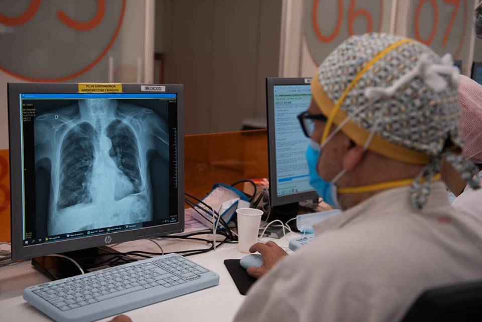
Simple chest X-ray. It is the first imaging test that is used in the diagnostic approach of patients with a suspicion of interstitial lung diseases. For that reason, a computed tomography (CT) scan of the chest is necessary to determine the type of disease.  

CT or MRI

Computed tomography (CT) scan of the chest. It is the main test to diagnose interstitial diseases. The different image patterns can suggest a particular disease, which, added to the clinical and laboratory characteristics can, in some cases, avoid the need to perform a lung biopsy. In the case of idiopathic pulmonary fibrosis, the characteristic radiology pattern is usual interstitial pneumonia (UIP). In the other interstitial diseases, the pattern can vary depending on the disease type. 

Patient performing spirometry

Simple spirometry, combined with plethysmography (lung volume measurements), and the carbon monoxide diffusing capacity test. They are of great use in the study of interstitial lung diseases. They confirm the diagnosis when they show restrictive type changes with a low carbon monoxide diffusing capacity. Similarly, they are used to assess the severity of the lung condition and for the follow-up of the patients. 

Person with electrodes at a running appointment performing a stress test

Six-minute walk test. It allows for the endurance capacity to be assessed in patients with pulmonary fibrosis.  

Person undergoing bronchoscopy

Fibre Bronchoscopy. In some cases, it is necessary to perform this type of procedure in order to take samples of the lower airway secretions (trachea, the bronchi and bronchioles, and the alveoli) and, occasionally to take biopsy samples.  

A person lying on a stretcher and a doctor performing a CT-guided lung puncture

Lung biopsy. When the clinical, radiological, and laboratory findings, and the other ancillary tests are unable to reach a consensus diagnosis, it makes it necessary to perform a lung biopsy using video-assisted thoracoscopy. The aim of the test is to obtain specimens of lung tissue so that it may be assessed by the multidisciplinary committee. 

Substantiated information by:

Fernanda Hernandez Gonzalez
Jacobo Sellarés Torres
Joel Francesqui
Sandra Cuerpo Cardeñosa
Xavier Alsina Restoy

Published: 9 June 2020
Updated: 9 June 2020

The donations that can be done through this webpage are exclusively for the benefit of Hospital Clínic of Barcelona through Fundació Clínic per a la Recerca Biomèdica and not for BBVA Foundation, entity that collaborates with the project of PortalClínic.

Subscribe

Receive the latest updates related to this content.

Thank you for subscribing!

If this is the first time you subscribe you will receive a confirmation email, check your inbox

An error occurred and we were unable to send your data, please try again later.