Diagnosis of Multiple Sclerosis

Reading time: 2 min

There are no tests that can establish the diagnosis of multiple sclerosis. Criteria are used —called McDonald criteria— which include:

  • Clinical manifestations indicative of a demyelination lesion.
  • Presence of lesions in magnetic resonance (MR) scan, which must meet a series of established characteristics.
  • Exclusion of other situations that could produce a similar clinical picture.

Taken together, the criteria help to define that there are two different areas of the central nervous system (CNS) involved and at two distinct times. That is to say, there is involvement in different areas of the CNS (dissemination in space) and at different times during the course of the disease (dissemination in time), which is what characterises the disease.

The criteria are established in order to diagnose those patients who have symptoms suggestive of demyelination, and, thus, in those in whom the disease is suspected, and not to differentiate the disease from other neurological disorders.

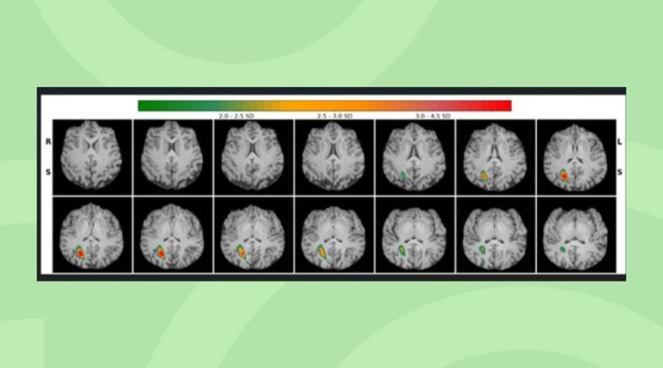
A clinical interview (anamnesis) is conducted with the patient in order to determine the symptoms and to check that the neurological changes respond to the lesions shown in the MR scan. The MR scan is useful to observe if there are multiple lesions, indicative of dissemination in space, and also in order to rule out other diseases or processes.

It is important that a neurologist—if possible  familiar with the disease—applies the criteria and makes the multiple sclerosis diagnosis.

5.2. Tests for Multiple Sclerosis

Magnetic Resonance Imaging or MRI machine

Magnetic Resonance (MR). A brain MR scan is performed on all patients and, in many cases, a spinal cord MR scan. It is advisable that the examination includes the administration of a contrast (gadolinium) in order to observe if there is evidence of dissemination in time, since recent lesions uptake contrast different to chronic ones, and helps in the differential diagnosis.

Evoked potentials

Evoked potentials. They register the electrical signals produced by the central nervous system (CNS) as a response to sensory stimuli (visual, electrical, auditory or tactile). This test helps to identify any silent involvement in different areas of the CNS (dissemination in space) or to confirm involvement at different times during the course of the disease (dissemination in time).

Bone marrow harvesting from the lumbar region

Lumbar puncture. A small sample is drawn from the cerebrospinal fluid (CSF) that protects and cushions the brain and the spinal cord. This analysis helps to determine, through the proteins called immunoglobulins that indicate inflammation in the CSF (oligoclonal bands), if there are anomalies in the antibodies, infections, and other diseases with similar symptoms to multiple sclerosis. This test is not necessary for the flare-up forms, but it is advisable for the primary progressive forms.


Blood collection tube

Blood analysis. They are more or less dependent on the clinical history of the patient, the neurological examination, and the results of the magnetic resonance scan.

Substantiated information by:

Albert Saiz Hinarejos
Ana Hernando Andrés

Published: 20 February 2018
Updated: 20 February 2018

The donations that can be done through this webpage are exclusively for the benefit of Hospital Clínic of Barcelona through Fundació Clínic per a la Recerca Biomèdica and not for BBVA Foundation, entity that collaborates with the project of PortalClínic.

Subscribe

Receive the latest updates related to this content.

Thank you for subscribing!

If this is the first time you subscribe you will receive a confirmation email, check your inbox

An error occurred and we were unable to send your data, please try again later.