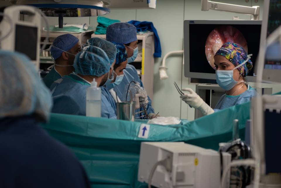
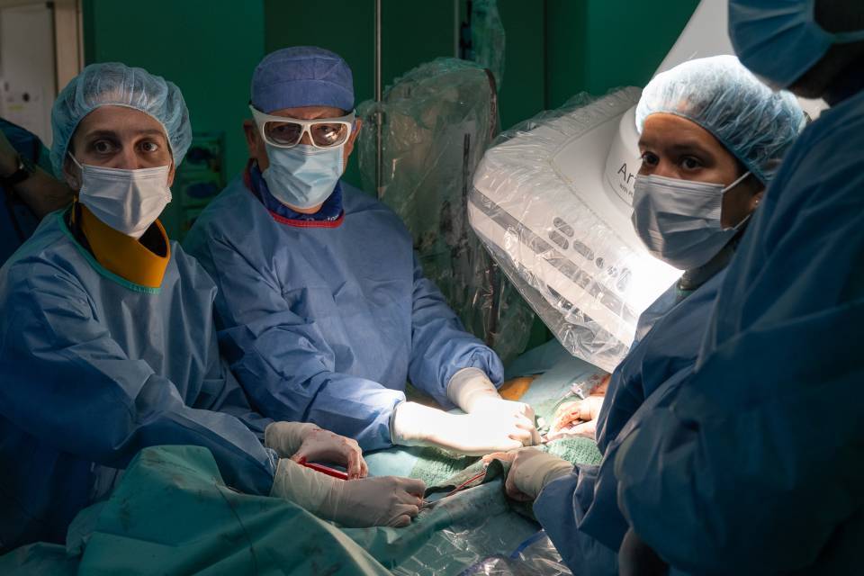
The Hospital Clínic carries out 50 skull base surgeries each year and is the only hospital in Spain to perform this technique. The description of the technique, used in very few centres worldwide, and the application in patients at the Hospital Clínic de Barcelona, has been published in the ‘Journal of Neurosurgery’. The Hospital Clínic had already performed 20 surgeries with this technique.
Transorbital neuroendoscopic surgery is an effective surgical technique for treating a variety of advanced brain diseases (especially in patients with skull base tumours). This type of minimally invasive surgery is performed through the eye socket, thereby avoiding the need to remove the top of the skull in order to access the brain. This avoids having to make a larger incision with all the possible associated complications. In some cases, skull base tumours can be extracted by adopting a combined endonasal and transorbital approach.
In order to achieve access, the surgeons make a small incision on the upper eyelid crease. A small hole is then made through the bone of the eye socket, measuring only two or three centimetres, to reach the brain. Surgery time is much shorter, since the skull does not need to be repaired and there is no need to close a large incision. This approach also allows repairs to be made without lifting the brain, protecting the optic and olfactory nerves as well as the carotid and ophthalmic arteries. Moreover, the advantages of this transorbital approach include reduced pain and decreased recovery time for the patient.
The transorbital neuroendoscopic surgery procedure has been used to repair cerebral spinal fluid leaks, for optic nerve decompression, to repair skull base fractures and eliminate tumours. The skull base is the area between the eyes and behind the nose all the way to the brain and the spinal cord. It is one of the most complex areas in the human body, since a large number of nerves and the major blood vessels of the brain, head and neck pass through it. Skull brain tumours are rare and affect fewer than 1/100,000 people per year. Histologically, they are a diverse group of tumours and potentially pose significant management problems due to their proximity to the orbit and the intracranial cavity.
This technique is particularly indicated for patients with a tumour in the central part of the skull base, but also the intracranial compartment in the lateral cavernous sinus, in the medial temporal lobe or in lateral tumours in the third cranial nerve. The team of professionals at the Hospital Clínic started testing this technique on human cadavers before putting it to practice on patients.
The Hospital Clínic de Barcelona transorbital surgery programme can be carried out thanks to funding from a grant awarded by the TV3 Foundation and a grant from the Health Research Fund (FIS).
According to Dr. Joaquim Enseñat, head of the Neurosurgery Service, “this technique is a clear example of an innovative project that we are carrying out at the Hospital Clínic de Barcelona. It allows us to be more efficient in surgery and provide greater benefits and recovery for patients”.
According to Dr. Isam Alobid, coordinator of the multidisciplinary skull base surgery group, explains that, "this innovation is minimally invasive surgery, which aims to remove the skull base tumour whilst respecting the vital structures and the normal physiology of the nose”.
Meanwhile, Dr. Sánchez España, oculoplastic surgeon at the Hospital Clínic Ophthalmology Institute, says that, “the orbit is a strategically located structure of the skull, since it is a bony corridor directly connected to the brain. It needs to be handled carefully in order to avoid any possible ophthalmological complications”.
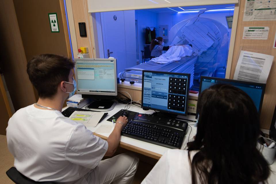
You can see a photo album of a transorbital surgery.


