The measurement of the proaggregating capacity of α-synuclein protein aggregates, (called seed) in cerebrospinal fluid through the seed amplification assay is a very robust diagnostic biomarker of Parkinson's disease. However, so far this assay has not been able to correctly distinguish between Parkinson's and multiple system atrophy, an atypical parkinsonism with a severity and prevalence similar to ALS but very difficult to clinically differentiate from Parkinson's in the early stages of the disease.
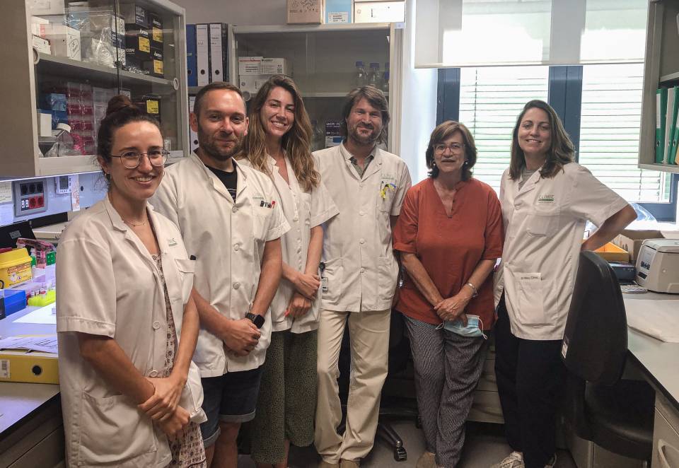
Now, a team from the Hospital Clínic-IDIBAPS has participated in a study published in the journal The Lancet Neurology that presents a modification of the seed amplification assay that is able to differentiate these two pathologies.
Yaroslau Compta, from the research group Parkinson's disease and other neurodegenerative movement disorders: clinical and experimental research of IDIBAPS, the Hospital Clínic Neurology Service, the Institute of Neurosciences UBneuro, the University of Barcelona, and one of the study’s authors, indicates: "Being able to differentiate these two disorders allows us to significantly shorten the diagnosis time, which is especially relevant for starting the appropriate treatment in time and ensuring an accurate prognosis of disability and survival". And he adds: "In addition, this modification of the seed amplification assay could allow us to include earlier cases in clinical trials of experimental therapies, a critical step for obtaining new treatments that slow the progression of what is an orphan disease in terms of treatments".
The study included 8 cohorts from 7 centres and 4 countries. In the most enriched cohorts in cases of multiple system atrophy, the new modified assay achieved a sensitivity of 87% and a specificity of 77%. Meanwhile, the two cohorts that had been obtained by applying stricter criteria, including that of the Clínic-IDIBAPS, had a sensitivity of 84% and a specificity of 87%.
Seed Amplification Assay
To perform the seed amplification assay, non-aggregated recombinant α-synuclein is added to biological samples (in this case cerebrospinal fluid) that are suspected of containing aggregates. The mechanical agitation of the sample causes the aggregates to fragment and interact with the non-aggregated recombinant protein, promoting a new aggregation. The aggregates resulting from the reaction are detected through a fluorescence reaction with thioflavin. This technique, in its original sensitive and specific modality for Parkinson's, is available at the Barcelona Hospital Clinic thanks to a project led by Dr Yaroslau Compta, with which the technique was developed at IDIBAPS.
In multisystem atrophy, α-synuclein is found in cytoplasmic aggregates in glial cells, while in Parkinson's disease it is found in Lewy bodies, located in neurons.
In the assay presented in this work, they observed that the aggregates of the Lewy bodies produced a very intense fluorescence. In contrast, the aggregates of the glial cells produced a positive reaction differentiable from the negative controls but with an intermediate fluorescence, which allowed the researchers to distinguish the two pathologies. Thus we speak of type 1 curves (for Lewy bodies such as Parkinson's and dementia with Lewy bodies) and type 2 curves in multisystem atrophy.


