IDIBAPS has several scientific platforms with specialist services for researchers of the institution and from other centers. One of these is the magnetic resonance imaging platform, which provides state-of-the-art techniques in magnetic resonance imaging.
A 3-tesla and a 7-tesla scanner
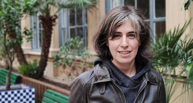
The platform’s story began in 2000 with the agreement signed between the Hospital Clínic Center for Diagnostic Imaging and the Universitat de Barcelona to acquire the first scanner. Since then, the platform has been constantly evolving, with the incorporation of new scanners, changes of location, and even the creation of an imaging laboratory. Much of this transformation has been due to the work of the team led by Núria Bargalló, who has been responsible for the platform for many years and is currently its scientific director. Now, a new team is facing the challenge of raising the profile of the platform’s work and becoming a gold-standard service for researchers.
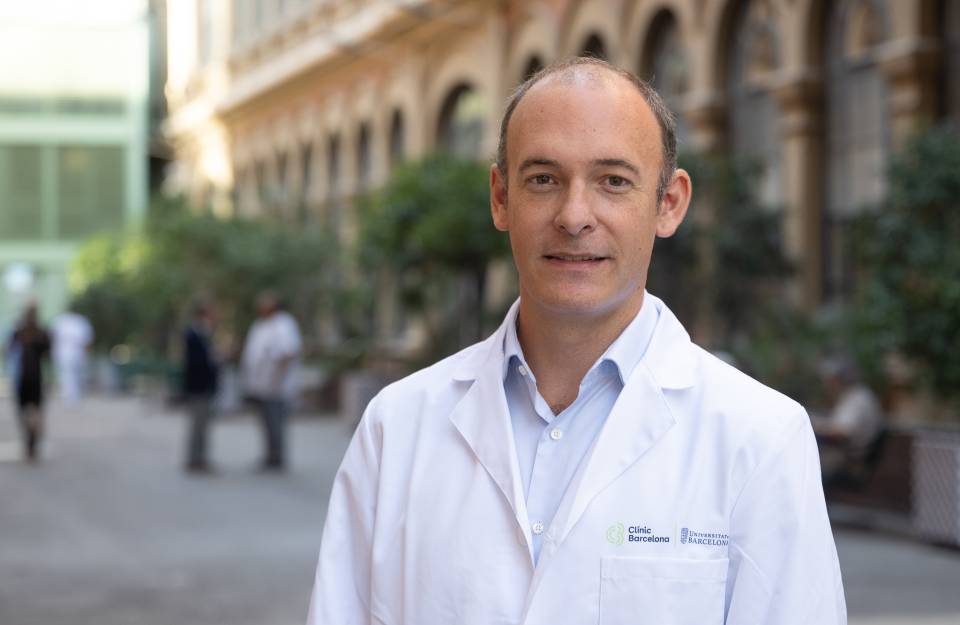
The platform currently has two scanners: a 2-tesla scanner for research in humans and a 7-tesla scanner for research in small laboratory animals and post-mortem tissue samples. “The tesla is the unit that indicates the power of the magnetic resonance instruments. In routine clinical practice, scanners of one-and-a-half teslas are used. Image resolution is sufficient for clinical diagnosis, but we need more detail in research”, explains Emma Muñoz-Moreno, head of the platform since last July. “The more teslas the device has, the better the resolution. This means we can distinguish finer and more sophisticated structures. In animals, we need scanners with a higher resolution than in humans, because their organs and tissues are smaller. We need to work at the micron scale rather than the millimeter scale. Increasing the power, however, also increases noise or distortion, which needs to be removed during image processing”.
Consulting on projects
The platform is much more than a service for acquiring magnetic resonance images. It also provides advice on project design, and the team is involved in writing and preparing the scientific articles produced. “Not all researchers are aware of the potential of magnetic resonance, beyond the standard applications. So, we accompany them from the outset, when they are looking at how imaging techniques can help them in their research, to the discussion of the results and the preparation of the articles. If they want, we can provide them with a comprehensive service”, explains Muñoz-Moreno.
Transforming images into information
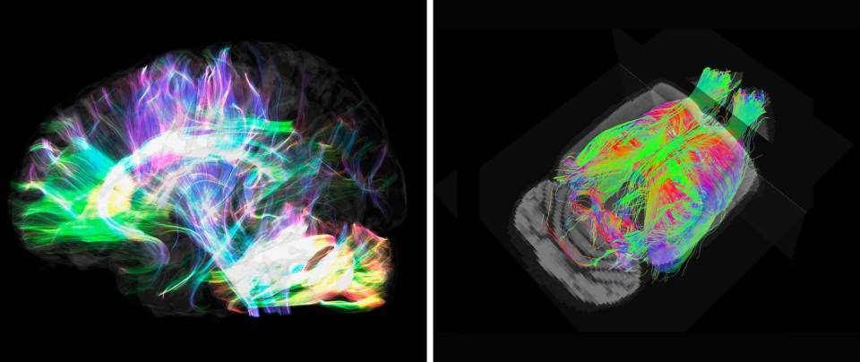
Using a powerful magnet and a sequence of radiofrequency pulses, magnetic resonance makes it possible to obtain a detailed view of the structure and properties of the body’s internal tissues. “It is important to note that the term ‘magnetic resonance’ includes different types of images. Depending on how we adjust the scanner parameters, we can obtain structural images, diffusion images, functional images, spectroscopy images, or angiography images, to name a few examples”. The images are in black and white, but it is the intensity of these colors that provides information on the different properties of the tissues. “For example, in the brain, the fact that an area is lighter or darker can allow us to differentiate the white matter from the gray matter in a structural image. In other types of image, the intensity can also be linked to the microstructure of the tissue, neuron activation, metabolite concentration, or blood flow. To obtain all this information, we use processing algorithms specific to each type of image”.
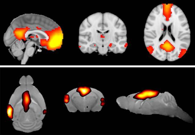
Functional connectivity in the human (top) and mouse (bottom) brain, obtained by resting-state functional magnetic resonance imaging. The colored areas correspond to the activation of the default neural network.
Actually, the platform makes a point of highlighting its image processing service. Muñoz-Moreno, a telecoms engineer, together with two biomedical engineers and two physicists, is responsible for carrying out the work of transforming the matrix of ones and zeros that make up the original black-and-white image into color images of the nerve fibers that connect the areas of the brain or of the neural networks that are activated while the subject carries out an action. “We design and apply the mathematical models for identifying, reconstructing in three dimensions, and quantifying the activation of the different areas of the brain”, says Muñoz-Moreno. There are few platforms that have teams or groups with the necessary knowledge to do this type of analysis. This is why the platform receives so many requests, even from researchers from other countries. “Today, image processing is the largest part of the platform’s work. The team also has technicians for the resonance devices, who are responsible for acquiring the images, and a biologist specializing in the care and handling of the animals, and they carry out essential work for the functioning of the service”.
Studying organs and tissues, even after death
A large part of the projects on which the platform collaborates involve neuroimaging. Of note is the research in psychiatry, in both children and adults, multiple sclerosis, stroke, Alzheimer, and Parkinson. For example, in the case of Parkinson disease, the researchers have studied how alpha-synuclein, the protein linked to the death of neurons in the black matter and the appearance of movement disorders, affects brain connectivity in mice. And another group has found differences in the structure of the brain stems of patients compared to those of healthy subjects.
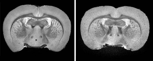
High-resolution structural image of the brain of Fisher rat (left) and TgF344-AD rat (right), a transgenic model of Alzheimer's disease. The presence of dots mainly in the cortex related to amyloid-beta peptide plaques is observed.
But magnetic resonance has applications in many other areas of research and, recently, the platform began a study in the field of cardiology. Another area that the team wants to promote is the study of post-mortem samples. “Our idea is to explore this option. To determine whether everything we see after death reproduces what occurs in the living body, with the idea of finding markers or identifying mechanisms and structures that may be of diagnostic or therapeutic utility”, says Muñoz-Moreno. “Furthermore, as the size of the samples is usually small, we use the 7-tesla scanner, which allows us to obtain higher-resolution images than those of studies carried out in living humans. We would like researchers to know about the possibility of carrying out these experiments to collaborate with them or even with the biobank. We believe that it would be of great interest and would add value to the research being carried out at the institution”.
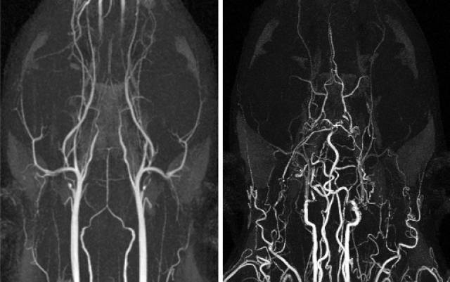
Vasculature in healthy rat brain (left) and after six months of bilateral carotid artery occlusion (right).
You will find more information and the contact details of the platform at https://www.clinicbarcelona.org/en/idibaps/core-facilities/magnetic-resonance-imaging
This content has been writen with the support of the Spanish Foundation for Science and Technology (FECYT).

