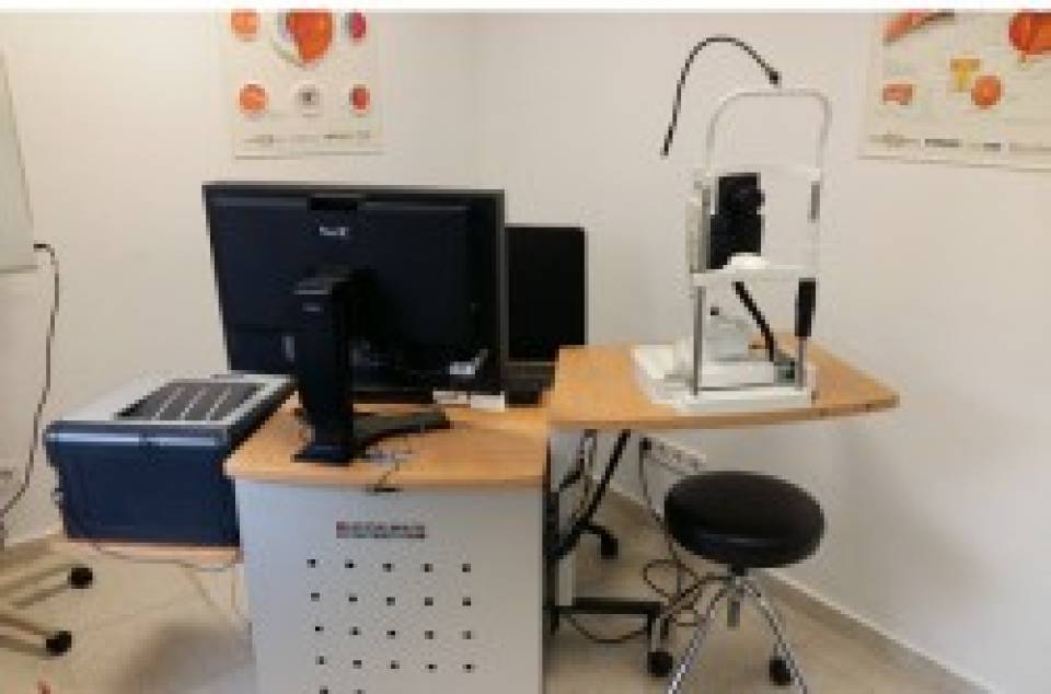
Multiple sclerosis (MS) is a disease that has an unpredictable course, so it is necessary an accurate patients’ follow-up to provide the best treatment. Development of imaging biomarkers for prediction of the clinical course of the disease would improve the management of patients. Most people with MS show inflammatory and neurodegenerative signs in the retina. Optical coherence tomography (OCT) is very reproducible and high-resolution imaging method for the assessment of retinal integrity.
In the study published in The Lancet Neurology, researchers included 879 patients with MS from 20 centers worldwide to assess whether the OCT is a useful technique to follow the course of the disease. This technique measured the thickness of the retina in patients over time, with a follow-up between 6 months and five years. They have established that below a certain retinal thickness, set at 88 microns, patients have a worse evolution of their illness. Thus, patients with retinal thickness below this value have twice the risk of disability worsening between the first and up to the third years of follow-up, and the risk increases uo to four times between 3 and 5 years of follow-up. The OCT is used to monitor disease progression and, because this technique less expensive and easier to perform than other used, as MRI, it can be useful for routine monitoring of patients. "The aim is not to replace the MRI, but OCT allows us to perform the test when the patient comes for consultation every 6 months", explains Dr. Elena H. Martinez-Lapiscina.
"Thanks to its high resolution, OCT can measure tiny changes that other techniques are impossible to identify. The idea is to incorporate this technique, used in ophthalmology, in the clinical practice in neurology. It could be also useful in diseases such as Alzheimer, Parkinson's disease or cerebral trauma", explains Dr. Pablo Villoslada. In addition, "our group is working on developing new technologies to monitor retinal and neurological diseases based on electrophysiology and molecular laser imaging, which would see the changes throughout the disease in an early stage" he concludes. For this purpose the IDIBAPS group collaborates with the Institute of Photonic Sciences (ICFO) and the Institute for Energy Research of Catalonia (IREC).
Article reference:
Martinez-Lapiscina EH, Arnow S, Wilson JA, Saidha S, Preiningerova JL, Oberwahrenbrock T, Brandt AU, Pablo LE, Guerrieri S, Gonzalez I, Outteryck O, Mueller AK, Albrecht P, Chan W, Lukas S, Balk LJ, Fraser C, Frederiksen JL, Resto J, Frohman T, Cordano C, Zubizarreta I, Andorra M, Sanchez-Dalmau B, Saiz A, Bermel R, Klistorner A, Petzold A, Schippling S, Costello F, Aktas O, Vermersch P, Oreja-Guevara C, Comi G, Leocani L, Garcia-Martin E, Paul F, Havrdova E, Frohman E, Balcer LJ, Green AJ, Calabresi PA, Villoslada P; IMSVISUAL consortium.
Lancet Neurol. 2016 Mar 18. pii: S1474-4422(16)00068-5. doi: 10.1016/S1474-4422(16)00068-5.

