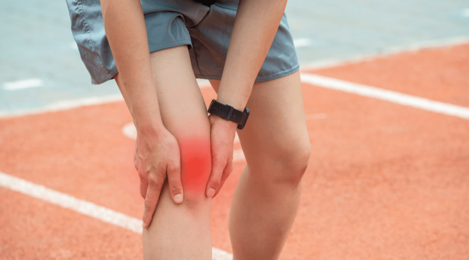
A research article, recently published in Proceedings of the National Academy of Sciences (the scientific journal of the US National Academy of Sciences), explains that a new biomaterial has been developed that regrows damaged cartilage in joints, a tissue that is very difficult to repair.
This research consisted of applying this biomaterial (which contains a biologically active protein, hyaluronic acid and other compounds) to the knee joints of sheep. In just 6 months, they found clear signs of repair and even growth of new, high-quality cartilage.
The knee structure, size and complexity in sheep closely resemble human knees, making them suitable for regeneration studies. If human trials prove successful, the study's authors believe this new material could be used to treat or potentially cure injuries affecting the hyaline cartilage in joints.
What are the current treatments for cartilage injuries?
Today, the different techniques to treat injured cartilage are generally classified as reparative, reconstructive or regenerative:
Reparative methods (perforations and microfractures) help the formation of new fibrocartilaginous tissue, facilitating the access of both blood vessels and the stem or progenitor cells from bone marrow. However, the fibrocartilage obtained in this case is different from the original hyaline cartilage.
Reconstructive methods seek to fill the defects using tissue grafts from the patient or a donor. These are known as mosaicplasties.
Finally, regenerative methods take advantage of bioengineering techniques to develop hyaline cartilage tissue using the latest advances in pluripotent cells.
Among these latter methods, products are used that attempt to resemble the structure of normal cartilage tissue. To obtain these products, Matrix-induced Autologous Chondrocyte Implantation (MACI) techniques are used, where cartilage cells are implanted on gels and grown in vitro in the laboratory.
Examples of these products are: CarGel®, CartiFill® and Chondro-Gide®. Most of these products produce type I or III collagen. However, type II collagen, which is characteristic of normal hyaline cartilage, has not yet been obtained.
The size and depth of the lesion, the level of activity of each person, the associated injuries to the meniscus and ligaments and the age of the patient are all factors that determine the appropriate technique for the treatment of the chondral lesion.
Why is cartilage so difficult to repair?
Articular cartilage is a highly elastic tissue with no nerves, blood or lymphatic vessels. It is designed as a lubricated surface to allow the bones to slide and rotate over each other with hardly any friction, throughout the joints. It reduces friction and distributes mechanical loads in different joint positions.
Articular cartilage has little or no capacity for regeneration by itself. Thus, wear and tear of the cartilage can cause osteoarthritis and functional deterioration in the medium and long term.
Repairing articular cartilage is a complex and difficult process due to the low cell density, the complex structural framework of the tissue and the inability of chondrocytes (specific cartilage cells) to migrate towards lesions to repair them.
In conclusion, the new technique described in this article, as well as regenerative techniques in general, are effective only in lesions of less than 2x2 cm surface area; and this is provided the subchondral bone is not affected. Thus, while it is an important advance in the treatment of cartilage lesions, this novel biomaterial cannot currently be used for advanced lesions; that is, in general osteoarthritis.
Furthermore, medium and long term clinical studies will need to establish whether the tissue obtained by these researchers is really hyaline cartilage or other types of collagen, as found in other cases. For now, therefore, the main treatment for joint injuries will continue to be joint prostheses.
Information documented by:
Dr Sergi Sastre, Traumatologist, Clinical Institute of Medical and Surgical Specialities.


