Heart attacks and other injuries to the heart leave scars in cardiac tissue that can disrupt the signals that regulate heartbeat circulation. This poor signal transmission can lead to ventricular tachycardia, a life-threatening arrhythmia and one of the leading causes of sudden death. Cardiac ablation is a procedure used to remove the areas that generate arrhythmias in this damaged tissue, thereby preventing abnormal electrical signals or rhythms. Now, a team at IDIBAPS-Hospital Clínic Barcelona has shown that magnetic resonance imaging (MRI) can help medical practitioners to evaluate the effectiveness of cardiac ablation and predict the risk of tachycardia in the future.
Published in the European Heart Journal: Cardiovascular Imaging, the study, PAM-VT, tracked the performance of MRIs on 49 patients after ablation. Its findings show that after ablation, there is a clear decrease in areas of cardiac tissue with low conductivity, which potentially cause tachycardia. The researchers also observed that the decrease in these areas correlates with less risk of suffering from tachycardia.
PAM-VT was led by Ivo Roca-Luque, the head of the Arrhythmias Section of Hospital Clínic Barcelona’s Cardiology Service and a member of the IDIBAPS research group Biopathology and treatment of cardiac arrhythmias. It was conducted in partnership with the Cardiac Imaging Section and the rest of the Cardiovascular Institute (ICCV). According to Roca-Luque, ‘We have shown that magnetic resonance imaging is a good tool for evaluating the effectiveness of ablation and for predicting the risk of future ventricular tachycardias. This procedure may allow us to classify patients based on their risk of recurrence to provide them with personalised and more effective monitoring’.
What we knew about the usefulness of this kind of magnetic resonance imaging
The cardiac magnetic resonance technique used in this study, called late gadolinium enhancement cardiac magnetic resonance (LGE-CMR), had previously proven effective in identifying areas of cardiac tissue that may involve abnormal signal transmission in studies conducted by the same group. However, its usefulness had always been demonstrated before and during ablation to help medical practitioners to facilitate and plan the procedure.
‘After many years of research on the role of magnetic resonance to predict arrhythmias in patients without ablation and to guide ablation, there were still no clinical data on whether it could identify injuries caused by the ablation itself and whether these data could be related to the risk of tachycardia recurrence’, Roca-Luque says. ‘That is why we identified the need for a study aimed at determining whether this magnetic resonance technique is useful for evaluating the effectiveness of ablation, comparing images before and after the procedure’.
New arrhythmia ward
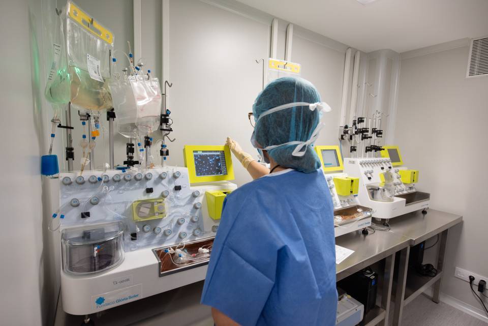
A few weeks ago, the newly renovated arrhythmia area, led by Ivo Roca-Luque. The new wardshave the best technology currently available for performing pacemaker and defibrillator implants and ablations (both simple and complex, such as ventricular tachycardia, etc.). The new facilities have the technology required for connections and training anywhere in the world.
Patients who undergo procedures in the new arrhythmia wards come from A&E, hospital wards, critical care units and day hospitals. The Clínic is the centre in Spain with the highest volume of complex ablations and the highest volume of physiological stimulation systems. In cardiology, physiological stimulation implants are devices used to regulate and improve cardiac function, mainly by means of electrical impulses. These devices are used to treat heart rhythm disorders and/or heart failure. The most common are: cardiac pacemakers and ICDs. Pacemakers are devices that control the heart rhythm in patients with bradycardia (slow heart rhythm) or heart block. They send electrical impulses to the heart to maintain a regular and adequate beat. Implantable cardioverter defibrillators (ICDs), meanwhile, are devices that monitor the heart rhythm and act when they detect serious arrhythmias such as ventricular tachycardia or ventricular fibrillation, and deliver electrical impulses to re-establish the normal rhythm (as well as functioning as pacemakers). Moreover, these patients with ventricular tachycardia undergo ventricular tachycardia ablation, with a comprehensive approach to planning (magnetic resonance imaging, external mapping systems, etc.) which allow the Hospital Clínic to be a world-class centre for these patients and for training in these techniques.
Seven doctors, seven professionals undergoing formal training, and nine nursing professionals work in this facility. The Arrhythmia Section performs around 400 complex ablations per year, 100 device implants, and monitors around 2,000 patients remotely. The ward has been refurbished and equipped with the latest technology thanks to financing from European Funds.


