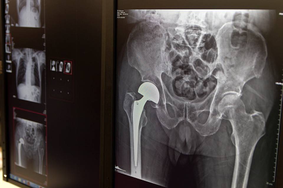
One of the most common complications in hip replacement surgery is infection. The main cause is the presence of areas of necrotic bone, i.e., zones where cell death has occurred, and where the tissue can no longer recover. For this reason, during hip replacement surgery, all non-viable tissue must always be removed. However, identifying this necrotic tissue is complex. Establishing the boundaries of the area to be removed is often dependent on the subjective judgement of the surgeon.
In this study, carried out by the Orthopaedic Surgery and Traumatology Department at Hospital Clínic in Barcelona, tetracyclines, a type of antibiotic, were used to more precisely define which areas of bone should be removed. This antibiotic was selected because it has a particularly useful feature: it fluoresces when irradiated with ultraviolet light. In addition, tetracyclines are deposited in the calcium of healthy bone, making it possible to differentiate between areas where necrosis has already occurred and those where it has not.
This technique is used in other fields, including maxillofacial surgery and traumatology, but it had never before been used in prosthetic surgery. To test whether the technique is effective in these cases, a preliminary study was conducted on three patients with chronic and severe hip prosthesis infections, treated with tetracyclines. During the prosthesis replacement operation, the area was irradiated with ultraviolet light to show where the bone fluoresced and thus define more precisely which areas of the femur were to be removed.
The bone that was taken out from all three patients was analysed anatomopathologically, and signs of bone infection were found in the areas that had been removed. One year after the surgery, none of the three patients had suffered further infection. These results suggest that all the infected or necrotic parts of the bone had been removed accurately and effectively.
The conclusion of the study is that using bone fluorescence after tetracycline marking is a useful tool for detecting the presence of non-viable bone. For this reason, the technique should be tested in more extensive studies to see if it could be applied more widely in hip replacement surgery.
Author: Dr. Ernesto Muñoz, Orthopaedic Surgery and Traumatology Department, Hospital Clínic, Barcelona.


