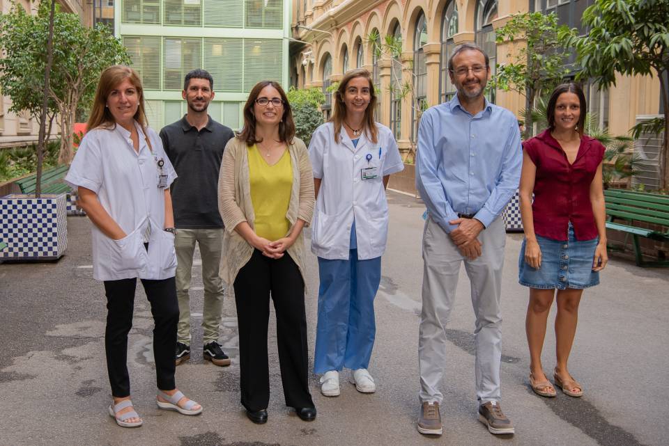
The study was led by Fàtima Crispi and Eduard Gratacós, of the Maternal-Fetal Medicine Department of BCNatal (Hospital Clínic and Hospital de Sant Joan de Déu) and the IDIBAPS Fetal and perinatal medicine group.
It also involved the participation, among others, of researchers from the Institut Clínic Cardiovascular, the Institut Clínic Respiratori, the IDIBAPS Cardiac imaging and Translational computation in cardiology research groups and researchers from the Universitat de Barcelona, Universitat Pompeu Fabra, the Barcelona Supercomputing Center and Philips Research France. The project also benefited from the support of the Fundació La Caixa, the Carlos III Health Institute, the European Commission, CEREBRA, CIBERER and AGAUR.
Low birth weight and cardiovascular risk
People who were born with low birth weight (those in the first decile, i.e. out of all births, the 10% of babies that were born with the lowest birth weight) experience more cardiovascular problems when they are adults. For example, they have up to three times more probabilities of suffering a myocardial infarction.
To date, the reason for this was unknown, and it was considered that it might be due to higher rates of heart attack, obesity, hypertension, stroke, diabetes or metabolic syndrome.
The research team led by Dr Gratacós was the first to demonstrate, in previous studies, that an important part of the problem is the heart itself. “We saw that the hearts of children with a low birth weight present differences in function and in structure, and that these differences that appear in the fetal stage persist until adolescence”.
It was still necessary to find out whether the changes in the heart’s structure and function persist in adult age and this was what was studied in the work published in JAMA Cardiology. “It is a pioneering study, which combines highly sophisticated computerised analysis techniques to analyse the shape of the heart using magnetic resonance imaging and a stress test”, explains Marta Sitges, director of the Institut Clínic Cardiovascular, head of the IDIBAPS Cardiac imaging group and co-author of the study.
People were selected aged between 20 and 40 years who had been born with low birth weight or with normal birth weight. To locate them, the delivery books from the Hospital de Sant Joan de Déu from 20 to 40 years ago were reviewed. Based on the date of birth and the mother’s surname, it was possible to contact some of the individuals, and the proposal was made to them to participate in the study.
Participation consisted of 158 adults, of whom 81 had been born with a low birth weight and 77 with a normal birth weight. They underwent a cardiac MRI and a stress test using a bicycle.
Differences in cardiac structure and response to stress
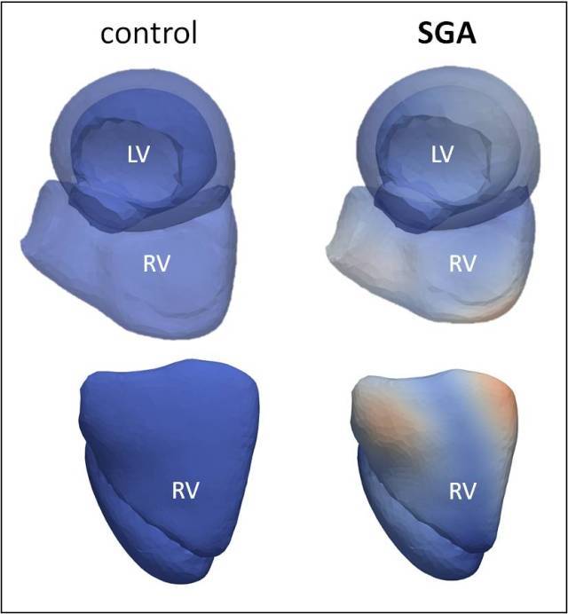
“The cardiac MRI showed that people who had been born with low birth weight show significant changes in theirr heart structure at adult age. Their right ventricle had a different shape”, explains Fàtima Crispi.

It was also seen that they had a lower capacity for exercise, in other words, they are not capable of generating as much workload with the bicycle and they become tired sooner. “That doesn’t mean that they can’t do exercise, to the contrary”, clarifies Crispi. “Simply, it may mean that they do not have the same capacity as the rest of the population and they get tired sooner”.
For Gratacós: “This research demonstrates again the importance of fetal medicine in the prevention of adult disorders. If we identify foetal growth problems during pregnancy and we promote healthy habits from childhood, we will avoid the consequences that foetal problems can lead to in adult life”, he concludes.
A recent study by the same research group has demonstrated that low birth weight multiplies by three the risk of suffering from severe COVID-19.


