Resting-state functional magnetic resonance imaging (rs-fMRI) is a non-invasive technique that enables the visualization of brain activation and identification of functional connectivity networks such as, for example, the motor network, the visual network, etc., when the subject is at rest. For this reason, this technique is widely used in clinical research in disciplines such as psychiatry or neurodegenerative diseases.
Its application to animal models has great translational potential, as it allows to obtain functional networks similar to those described in humans. However, to date there was no consensus protocol. “Different studies had been published involving resting-state fMRI in rats, but each research group implemented its own image acquisition protocol based on previous literature and incorporating its own modifications, which may difficult the comparison between studies", explains Emma Muñoz-Moreno, head of the Magnetic Resonance Imaging Core Facility.
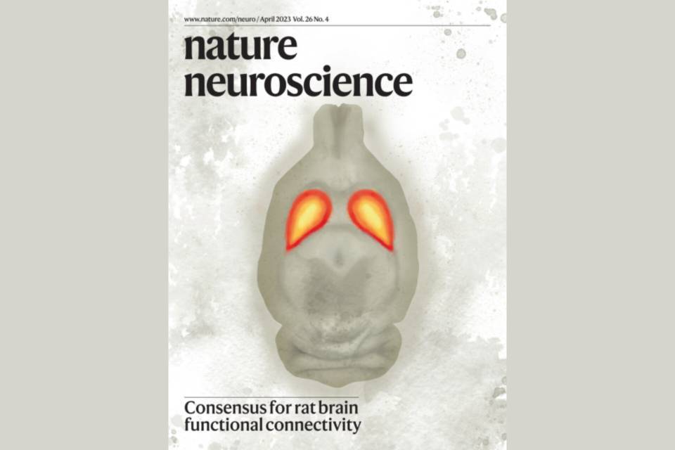
The aim of the study published in Nature Neuroscience was to define a common protocol for acquiring rs-fMRI data to optimise the identification of functional networks. For this, it started with a phase called MultiRat in which 46 centres provided data from previous projects to identify the parameters that optimise the identification of these networks. IDIBAPS contributed with acquisitions made within the framework of two projects led by Guadalupe Soria, that were focused on changes in connectivity in Alzheimer's Disease models.
From here, the consensus protocol was defined. It includes the parameters for the magnetic resonance image acquisition and the animal sedation . “Furthermore, since raw data from the scanner need to be processed via different algorithms to ensure their quality and extract the information of interest, the processing pipeline is also available," adds Muñoz-Moreno.
Subsequently, a second phase was carried out, called StandardRat, in which 20 centres took part to evaluate the consensus protocol. In this case, IDIBAPS provided the images obtained from 10 animals (5 males and 5 females) scanned with this protocol.


